With the help of radiographs (the proper term for pictures taken with X-rays), your dentist can look at what is happening beneath the visible oral tissues. Dental X-ray examinations provide valuable information that your dentist could not collect otherwise. They pose a far smaller risk than many undetected and untreated dental problems. If you have questions about your dental X-ray exam, talk with your Ottawa family dentist.
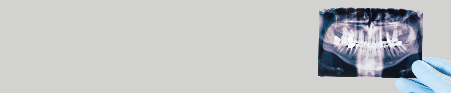
When X-rays pass through your mouth during a dental exam, more X-rays are absorbed by the denser parts (such as teeth and bone) than by soft tissues (such as cheeks and gums) before striking the film. This creates an image called a radiograph. Teeth appear lighter because fewer X-rays penetrate to reach the film. Tooth decay, infections and signs of gum disease, including changes in the bone and ligaments holding teeth in place, appear darker because of more X-ray penetration. Dental restorations (fillings, crowns) may appear lighter or darker, depending on the type of material used for the restoration. The interpretation of these radiographs allows your Ottawa family dentist to safely and accurately detect hidden abnormalities.
How often X-rays (radiographs) should be taken depends on the patient’s individual health needs. It is important to recognize that just as each patient is different form the next, so should the scheduling of X-ray exams be individualized for each patient. Your Ottawa family dentist will review your history, examine your mouth and then decide whether you need radiographs and what type. If you are a new patient, the dentist may recommend radiographs to determine the present status of the hidden areas of your mouth and to help analyze changes that may occur later. If you have had recent radiographs at your previous dentist, your new dentist may ask you to have the radiographs forwarded.
The schedule for needing radiographs at recall visits varies according to your age, risk for disease and signs and symptoms. Recent films may be needed to detect new cavities, or to determine the status of gum disease or for evaluation of growth and development. Children may need X-rays more often than adults. This is because their teeth and jaws are still developing and because their teeth are more likely to be affected by tooth decay than those of adults.
Many diseases of the teeth and surrounding tissues cannot be seen when your Ottawa family dentist examines your mouth. An X-ray examination may reveal:
Finding and treating dental problems at an early stage can save time, money and unnecessary discomfort. It can detect damage to oral structures not visible during a regular exam. If you have a hidden tumor, radiographs may even help save your life.
We are exposed to radiation every day from various sources, such as frequent airplane travel and high altitudes, minerals in the soil, and appliances in our homes (like smoke detectors and television screens).
| Source | Estimated Exposure (mSV*) |
| Dental radiographs x Bitewings (4 films) Full-mouth series (about 19 films) | 0.038 0.150 |
| Medical radiographs x Lower GI series Upper GI series Chest | 4.060 2.440 0.080 |
| Average radiation from outer space In Denver, CO (per year) | 0.510 |
| Average radiation in the U.S. from Natural sources (per year) | 3.000 |
Source: Adapted from Frederiksen NL. X-Rays: What is the Risk? Texas Dental Journal. 1995;112(2):68-72
*A millisievert (mSV) is a unit of measure that allows for some comparison between radiation sources that expose the entire body (such as natural background radiation) and those that only expose a portion of the body (such as radiographs).
A radiograph may be needed for dental treatment or a dental emergency that can’t wait until after the baby is born. Untreated dental infections can pose a risk to the fetus, and dental treatment may be necessary to maintain the health of the mother and child. Radiation from dental X-rays is extremely low. However, every precaution is taken to minimize radiation exposure. A leaded apron minimizes exposure to the abdomen and should be used when any dental radiograph is taken. A leaded thyroid collar can protect the thyroid from radiation, and should be used whenever possible. The use of a leaded thyroid collar is strongly recommended for women of childbearing age, pregnant women and children. Dental radiographs are not contraindicated if one is trying to become pregnant or is breast feeding.